MIZTLI Basic Tutorial
Last update: 2025-06-25
Foreword
This software is FOR RESEARCH USE ONLY! DON'T USE FOR DIAGNOSTIC PURPOSES!
25 June 2025: This tutorial was written 3 years ago and is now slightly outdated. However it remains a useful resource to grasp MIZTLI main features.
To use the software, navigate to miztli.biokerden.eu. You will need to use a recent web browser.
About This Tutorial
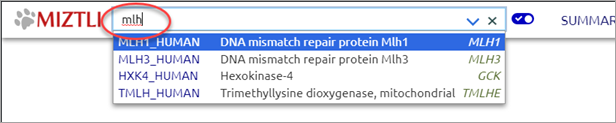
In this tutorial we will investigate a missense variant on human gene MLH1: Thr82Ala (NM_000249.4:c.244A>G) interpreted as Likely pathogenic in ClinVar.
Open MLH1_HUMAN
Go to miztli.biokerden.eu.
In the input field at the top of the page, start typing 'mlh' and then select MLH1_HUMAN (the related gene name is displayed on the right side of the list in italics).
Open Sequence
Click on 'SEQUENCE' in the top bar:
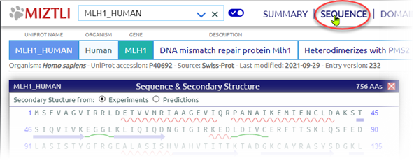
The MIZTLI user interface displays window-like panels inside the web page. You can move panels around, resize them, and close them at will.
The sequence panel displays the UniProt main isoform along with secondary structures.
Open Domains
Click on 'DOMAINS' in the top bar.
Hover the cursor over Thr82 in the Sequence panel. The amino acid position gets highlighted in the domains panel:
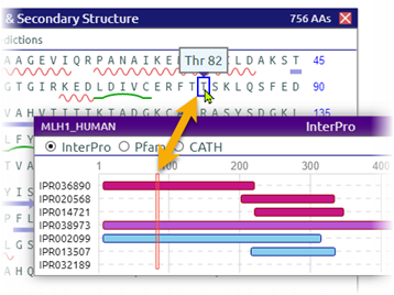
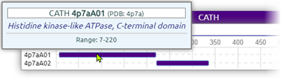
In the domains panel check the 'CATH' radio button. Hover the cursor over the domain including our variant: You can see that this domain has an ATPase activity.
Open Structures
Click on 'STRUCTURES' in the top bar.
This panel shows the known 3D structures available for the protein at hand (including experimental structures from PDB and AlphaFold-predicted structures).
Hover over the first segment of structure 4p7a. You can see that it includes position Thr82 (most experimental structures don't cover the full sequence).
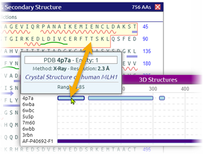
Right-click on the '4p7a' label to display details about the experimental method, resolution, and molecules included:
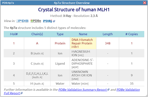
Beside a 348 amino acid segment of protein MLH1, this entry includes one Mg ion, one ADP molecule, a few unidentified atoms, and water.
Open a 3D View
MIZTLI integrates the Mol* (/'molstar/) molecule viewer.
Left-click on the '4p7a' label in the Structures panel. This opens Mol* showing the 4p7a structure:
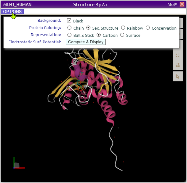
Mol* is very sophisticated but can be hard to use in the first place. This is why MIZTLI provides functional shortcuts and customized features. Click on the OPTIONS button as shown above to overlay several display settings.
To navigate in 3D space, use your mouse (Left-click: rotation - Ctrl-Left-click: translation, Wheel: scale). See Moving in 3D.
(The documentation of Mol* itself is available here. To get access to most Mol* settings, click on the ![]() icon.)
icon.)
Investigate AA Thr82 in 3D
In the 3D panel Options box, check:
- In the Protein Coloring field: Conservation
- In the Representation field: Surface
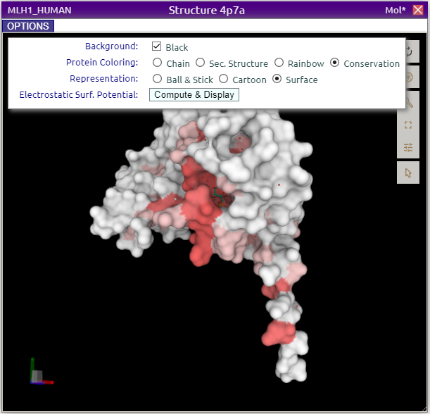
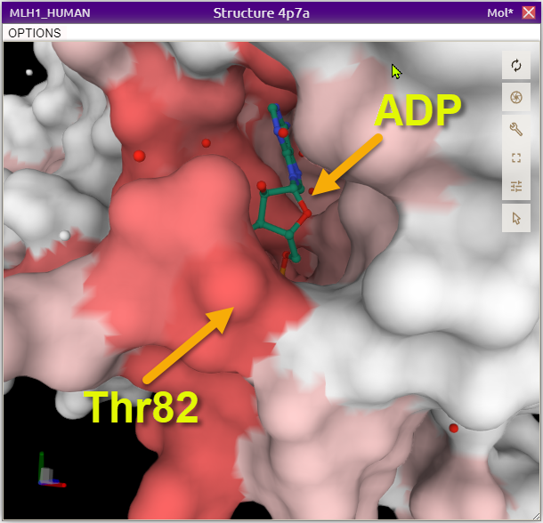
Rotate the view so as to identify the most conserved regions (the red ones).
Now, let's find where Thr82 is located in the 3D view. In the Sequence panel, hover the cursor over this amino acid to highlight it in the 3D view:

Thr82 is located in a conserved region, as seen graphically and textually at the bottom-right corner of the 3D view.
Zoom in on that region. You will see an ADP molecule in a pocket nearby:
Investigate Variant Thr82Ala
So, Thr82 is located in a conserved region, and it is close to the binding site of an ADP molecule.
We will now try to learn more about the possible effects of this Thr→Ala substitution. Here we will try to accurately predict how an Alanine sidechain fits in place of a Threonine at position 82 (keeping the backbone unchanged). This is called repacking and we will use FASPR for that purpose.
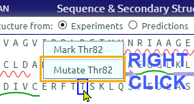
Let's 'create' variant Thr82Ala in MIZTLI.
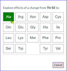
In the Sequence panel right-click on Thr82, select 'Mutate Thr82', and then select Ala.
![]()
This creates a variant panel as follows:
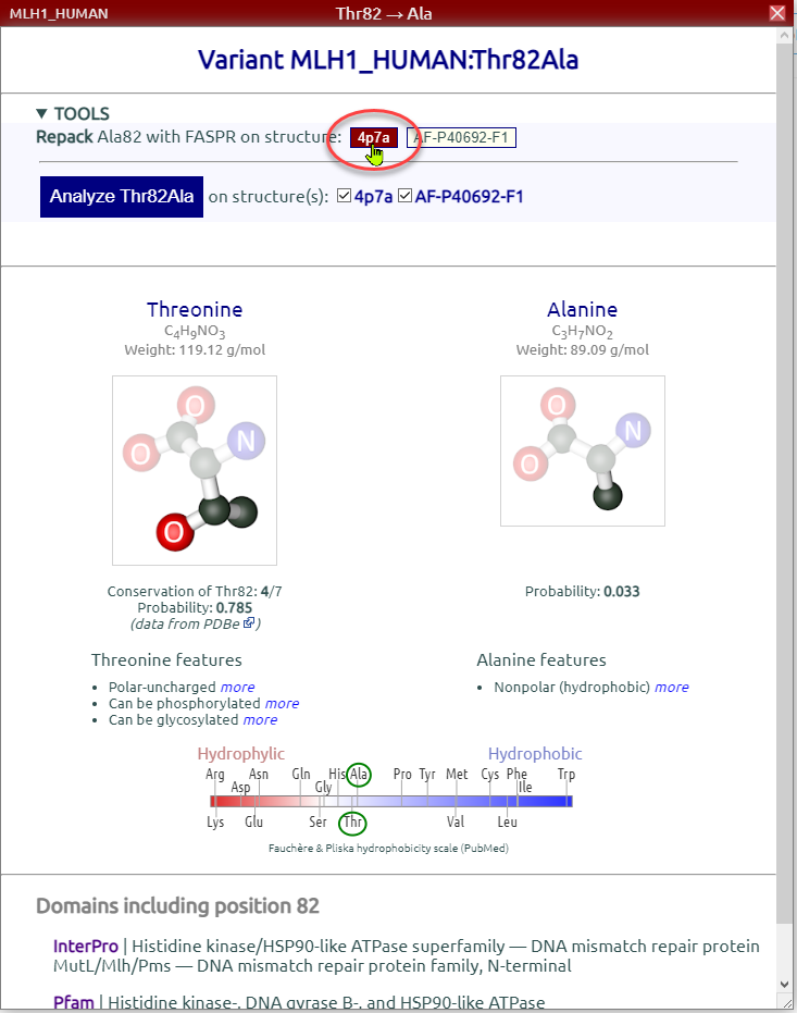
We will now replace Thr82 with Ala using FASPR on structure 4p7a (we could also use the AlphaFold predicted structure, AF-P40692-F1, but it does not include ligands).
In the variant panel click the 4p7a repacking button. We get a new 3D view focused on position 82 and its surroundings. In the Options sub-panel we can switch from Thr (REF) and repacked Ala (VAR).
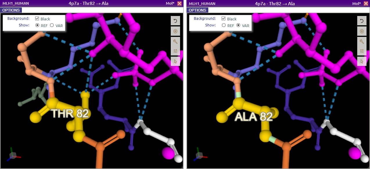
The magenta molecule here is ADP. Dashed lines represent noncovalent interactions between molecules such as hydrogen bonds. Moving around in the REF structure we can see that Thr82 interacts with ADP through two hydrogen bonds, and with Lys84 through one bond. These bonds are not found in the VAR structure. They might be important for the protein stability and even more for its ATPase activity.
Next Steps
- Let MIZTLI compute and report some of the potentially damaging structural effects of variants
- See how to display proteins' electrostatic surface potential
- Learn a bit more on how to move around in Mol* 3D representations
References
Sehnal, D., et al. (2021). "Mol* Viewer: modern web app for 3D visualization and analysis of large biomolecular structures." Nucleic Acids Res.
Huang, X., et al. (2020). "FASPR: an open-source tool for fast and accurate protein side-chain packing." Bioinformatics 36(12): 3758-3765.